Cardiac and cardiovascular image analysis has been performed at IIBI since the early 1990's, from X-ray coronary angiography, 2D/3D/4D ultrasound (including intravascular), MR/CT, and OCT, leading to established and validated software for various clinical questions and applications (coronary artery disease, aortic atherosclerosis, arrhythmia, heart transplant, etc.).
Cardiovascular image analysis at IIBI:
Sonka and Wahle Team

Milan Sonka
IIBI Co-Director
Andreas Wahle
Associate Research Engineer, Electrical and Computer Engineering
- Cardiac image analysis
- 3D/4D LV/RV analysis of MR images
- 3D/4D LV analysis of ultrasound images
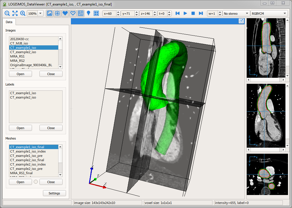
- Aortic image analysis
- 3D/4D analysis of aortic MR, MRA, CT, CTA
- Commercialization by Medical Imaging Applications, LLC
- Coronary image analysis
- Quantitative analysis of coronary angiography
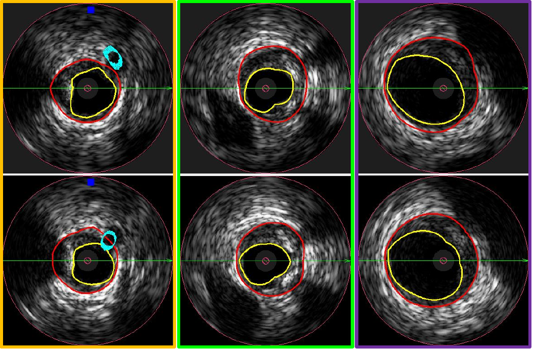
- Quantitative analysis of coronary intravascular ultrasound (IVUS)
- Geometrically correct analysis of coronary morphology from IVUS and angiography imaging
- Automated registration of IVUS image sequences
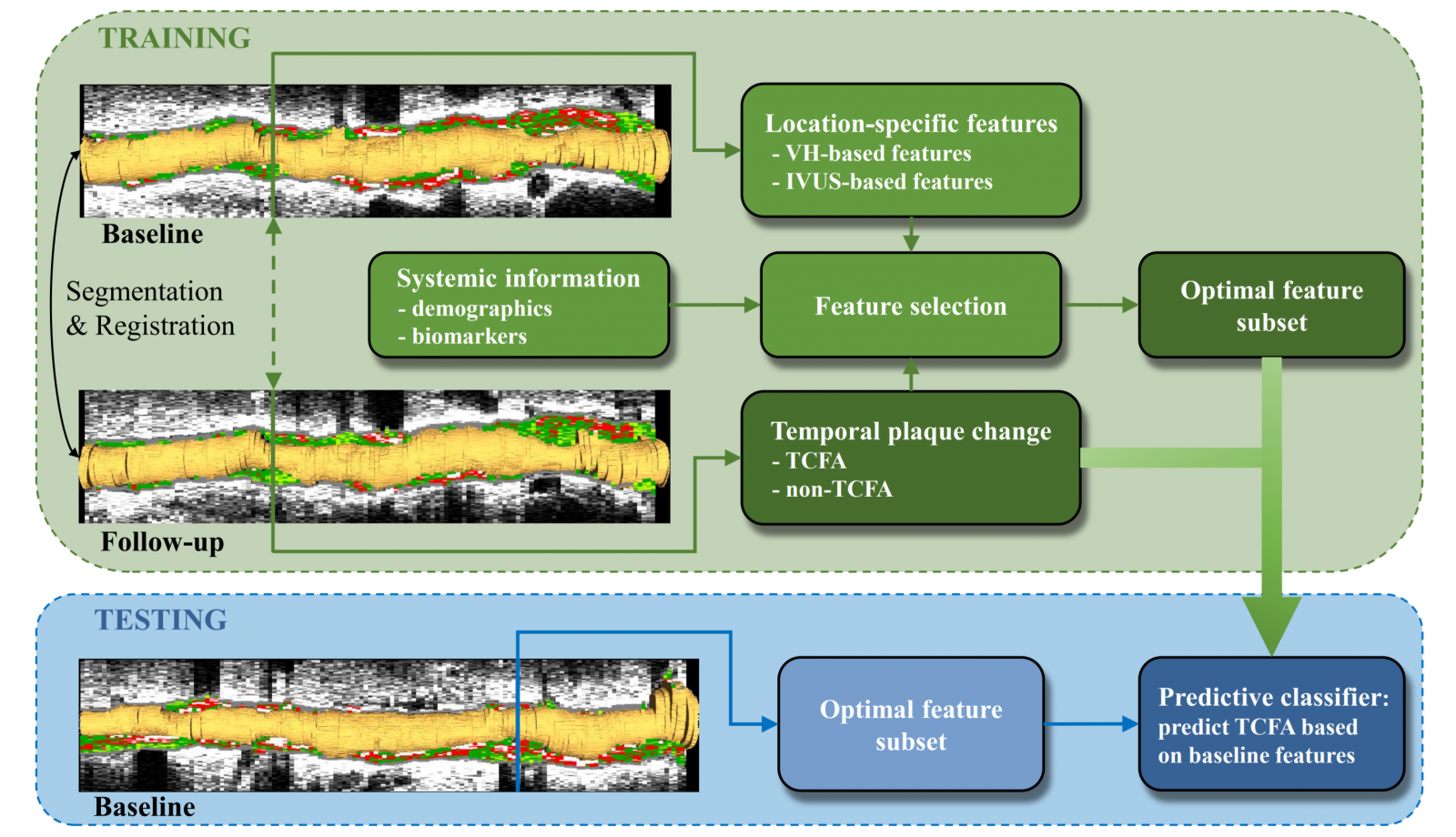
- Prediction of plaque progression from IVUS and angiography imaging
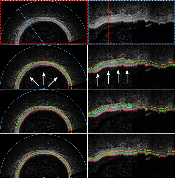
- Quantitative analysis of coronary OCT (optical coherence tomography)
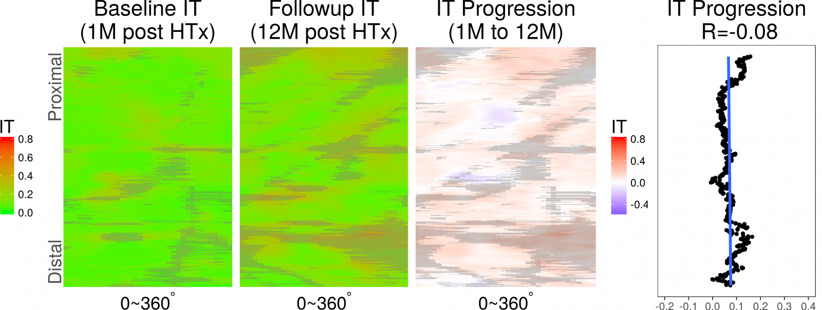
- Prediction of cardiac allograft vasculopathy from coronary OCT in heart transplant patients
- Publications
Cardiac image analysis
Left and right ventricles: 4D shape analysis and segmentation.
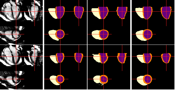
Left-ventricular segmentation from 3DRTE (3D real-time Echocardiography).

Aortic image analysis
3D/4D analysis of aortic morphology and wall calcification from CT. CTA, MR, MRA.
Coronary image analysis
Coronary OCT morphology analysis - color coded intimal-layer thickness. and its changes over time.

Coronary IVUS image segmentation in 3D pullbacks
Coronary IVUS image registration between 1-month and 12-month pullbacks
Thin-cap fibroatheroma prediction based on IVUS-VH