Current IIBI research investigates the pathology of many eye diseases, including diabetic retinopathy, macular degeneration, macular holes, epiretinal membranes, macular edema, central serous choroidopathy, and of the optic disc, combining state of the art imaging modalities like optical coherence tomography with our well-established segmentation approaches.
Ophthalmic image analysis is performed by the following mutually collaborating groups at IIBI:
Abrámoff, Kwon, Sonka Team

Michael D. Abramoff
Professor of Ophthalmology and Visual Sciences

Young H. Kwon
Professor of Ophthalmology and Visual Sciences

Milan Sonka
IIBI Co-Director
- Fundus image analysis
- Retinal abnormalities from fundus images
- Vasculature analysis from fundus images
- Commercialization by IDx
- Retinal OCT image analysis:
- Age-related Macular Degeneration, image-guided AMD treatment, prediction of AMD treatment outcome
- Glaucoma - Assessing visual function from 3D OCT
- Development of general-purpose OCT image analysis tools - Iowa Reference Algorithms
- OCT Explorer
- Just-Enough Interaction for multi-layer OCT image analysis
- Batch processing of tens of thousands 3D OCT image datasets
- Overarching publications
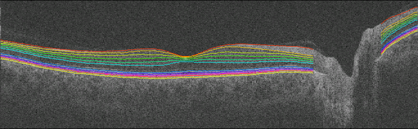
Retinal image analysis of 3D OCT - Iowa Reference Algorithms

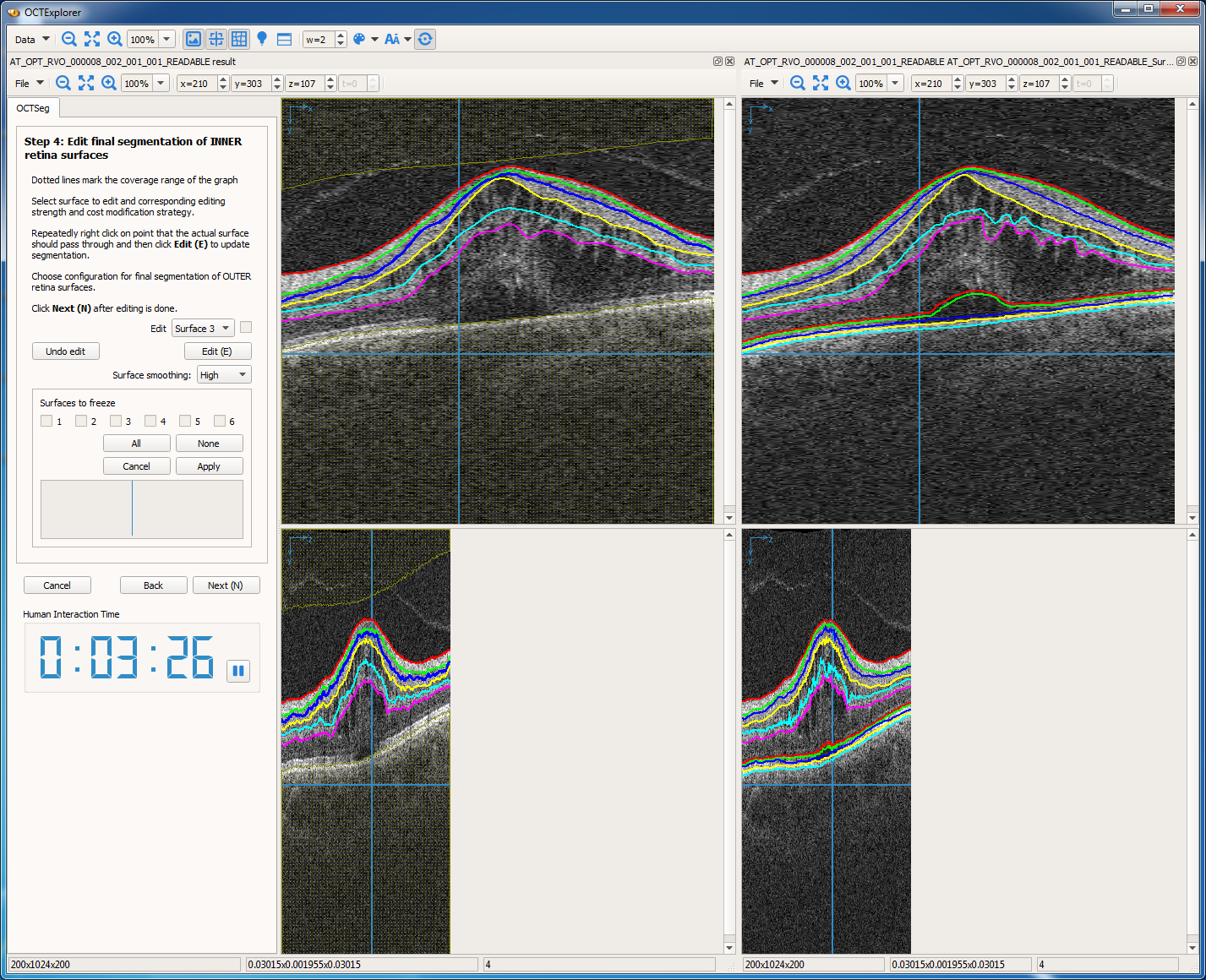
Left-to-right: OCTExplorer 3.8.0 (stable); OCTExplorer 5.0.0 (beta).
Choroid analysis from 3D OCT


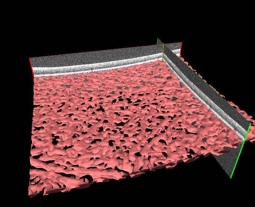
3D choroidal vessel segmentation. Left-to-right: Central B-scan (XY) image; en-face (XZ) image; 3D rendering.
Glaucoma - Assessing visual function from 3D OCT
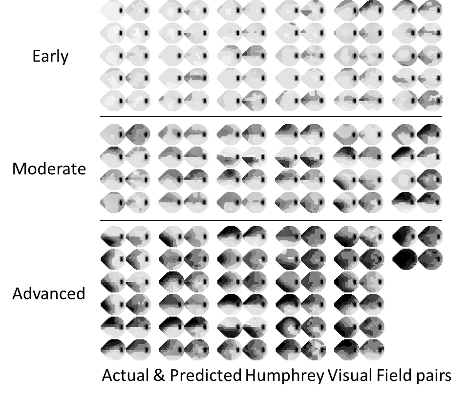
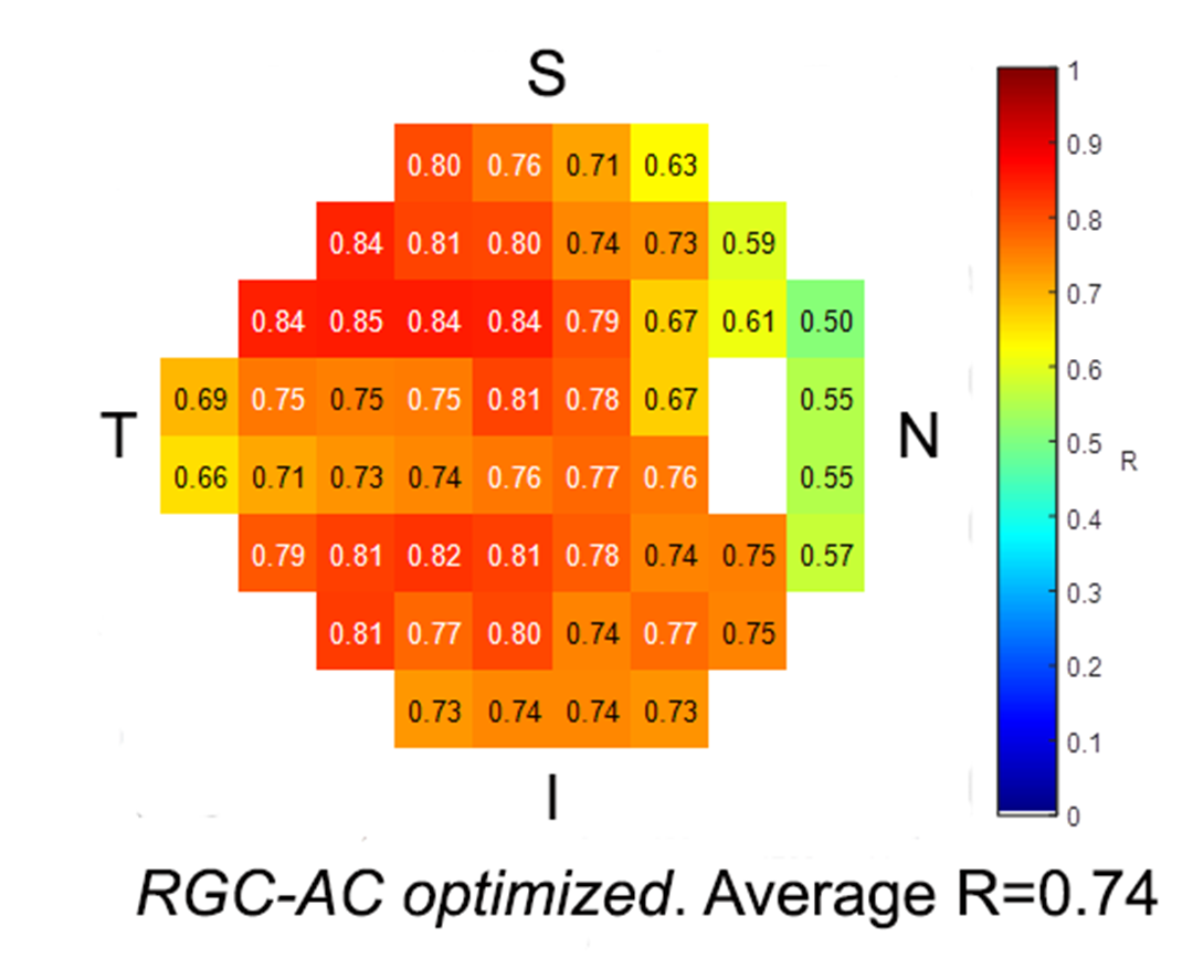

Left: actual and predicted 24-2 Humphrey visual field (HVF) pairs for 97 patients with early, morderate or advanced primary and secondry open angle glaucoma. Middle: Correlation of actual and predicted HVF at each test location, except locations at ONH, with predictors for each test location optimized based on retinal ganglion cell axonal complex (RGC-AC). Right: Optimized NFL path (green) for each predicted test location (red).
Age-related Macular Degeneration from 3D OCT
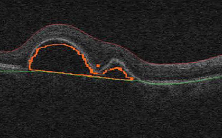
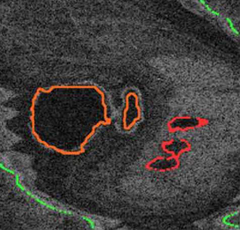


3D symptomatic exudate-associated derangements (SEADs) segmentation. Left-to-right: Central B-scan (XY) image; en-face (XZ) image; 3D rendering of retinal surfaces and SEADs; 3D rendering of SEADs.
ONH analysis from 3D OCT
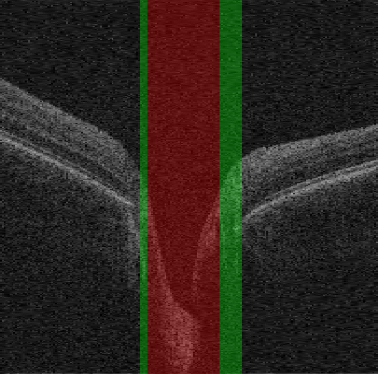
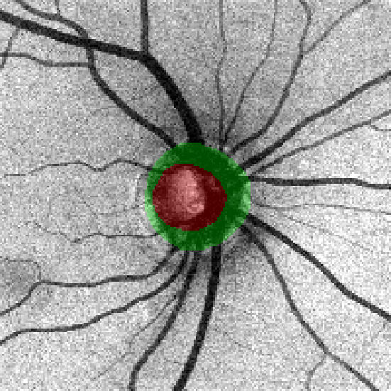

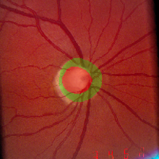
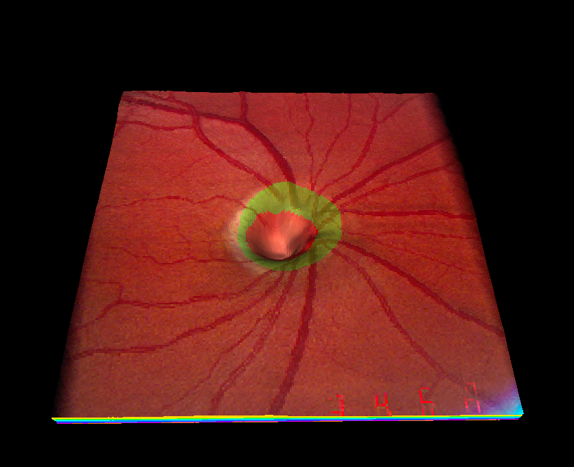
Optic disc segmentation (red: optic cup, green: neuroretinal rim). Left-to-right: Central B-scan image; OCT projection image; 3D rendering of the retinal surfaces textured using OCT projection image; registered fundus image; 3D rendering of the retinal surfaces textured using registered fundus image.
Garvin, Kardon Team

Mona Garvin
Professor, Electrical and Computer Engineering

Randy H. Kardon
Professor of Ophthalmology and Visual Sciences
- Peripapillary OCT image analysis
- Optic nerve head
- Papillodema
The University of Iowa Institute for Vision Research
Genetic aspects of ophthalmic diseases