Osteoarthritis and osteoporosis image analysis research is yet another focus area of IIBI. Architecture and biomechanics of osteoporotic bone, cartilage morphometrics, and overall orthopedic biomechanics are studies from a variety of imaging modalities, including MR, CT, and ultrasound.
Orthopedic image analysis at IIBI:
Saha Team

Pranav Saha
Professor, Electrical and Computer Engineering
- Projects
- Characterization of trabecular bone plate-rod micro-architecture
- CT-Based Modeling of Bone Micro-Architecture and Fracture-Risk
- Osteoporosis in Chronic Obstructive Pulmonary Disease (COPD)
- understanding the linkage between COPD-related factors, bone structure, and fracture risk and identification of unique COPD subgroups with elevated fracture-risk
- Publications
Andersen Team

Donald D. Anderson
Professor of Orthopedics and Rehabilitation
Sonka Team

Milan Sonka
IIBI Co-Director
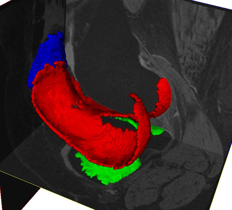
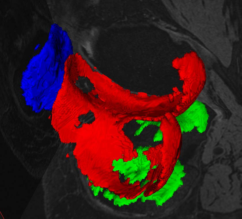
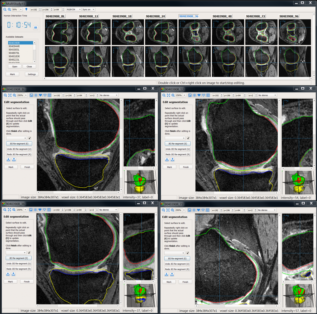
Cartilage image analysis inj the knee joint is of primary interest although the developed methods and approaches are generally applicable.
- Publications
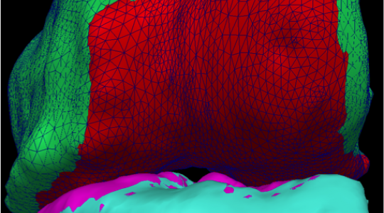
3D cartilage MR segmentation -- normal and osteoarthritic joints, note cartilage thinning and ``holes'' in the right-most image. Right panel: Simultaneous 4D knee MR bone+cartilage segmentation - 8 time-point 3D datasets registered and simultaneously segmented in 4D.