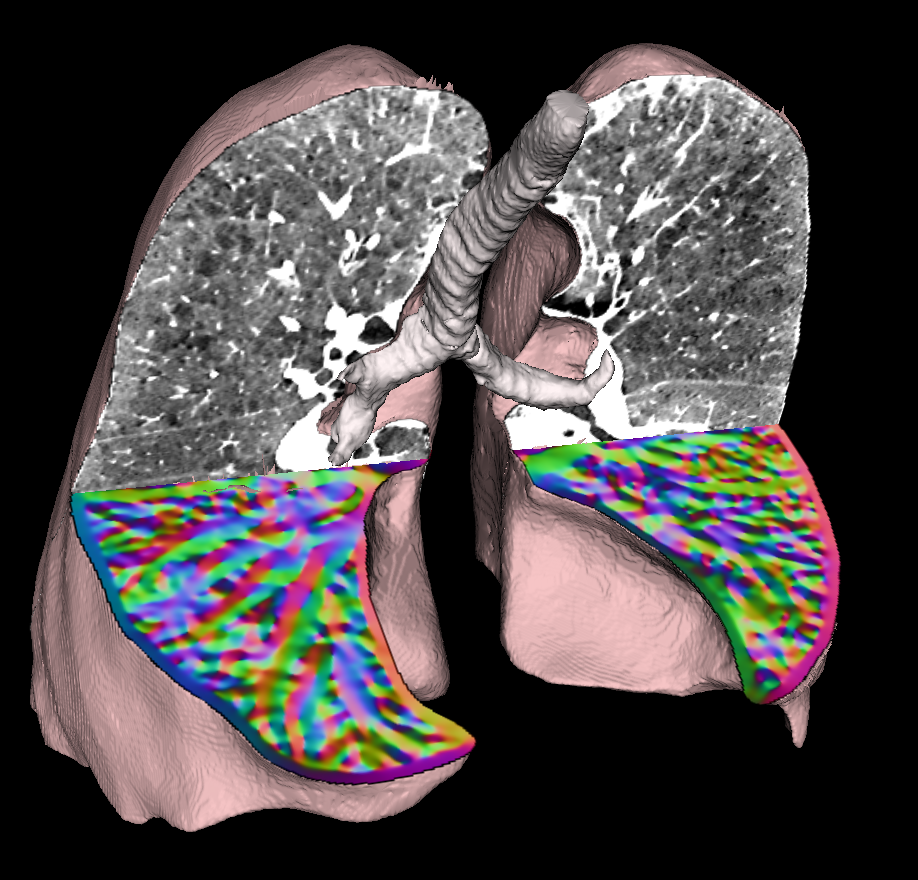
Development of novel imaging protocols for and image-related analysis approaches to assessing pulmonary morphology and function in normal lungs and various lung diseases is one of IIBI's strongest focus areas, going back 30+ years of research activity in various laboratories.
Lung image analysis at IIBI:
Hoffman Team

Eric A. Hoffman
Professor of Radiology-Division of Physiologic Imaging
- Advanced Pulmonary Physiomic Imaging Laboratory
- National Emphysema Treatment Trial (NETT)
- Severe Asthma Research Program (SARP), MESA-Lung, COPDGene
- Radiology Center, SubPopulations and InteRemediate Outcome Measures in COPD Study (SPIROMICS)
- https://appil.medicine.uiowa.edu/
Reinhardt Team

Joseph M. Reinhardt
Professor and Department Executive Officer, Biomedical Engineering
- Biomedical Imaging Lab (BME)
- Structural and functional evaluation of the normal and abnormal lung
- Lung tissue functional assessment to guide radiation therapy
- Analysis of breathing sounds to predict sputum accumulation during mechanical ventilation
- Pediatric airway segmentation, measurement, and shape modeling
- http://spect.ecn.uiowa.edu/
Christensen Team

Gary E. Christensen
Professor, Electrical and Computer Engineering
- Registration
- Deformable shape models
- Anatomical atlases
Beichel Team

Reinhard R. Beichel
Professor, Electrical and Computer Engineering
- Medical Computer Vision & Graphics Lab
- Quantitative lung image analysis in 3D and 4D CT scans
- Robust model-based segmentation of lungs with disease
- Quantitative lung image analysis in small animals (imaging cryomicrotome, micro-CT, etc.)
- Anatomically derived airway models and particle deposition data to facilitate computational toxicology in mice
- http://user.engineering.uiowa.edu/~rbeichel/
- Publications